lab values nursing mnemonics cheat sheets

lab values nursing
lab values nursing mnemonics
lab values
lab values nursing cheat sheets

lab values and what they mean

Laboratory Values and Interpretation – A Nurse’s Ultimate Guide
Getting familiar with and interpreting laboratory values can be intimidating, particularly to new nurses.
Reference Ranges and What They Mean
Laboratory test results reported as numbers are not meaningful by themselves. Their meaning comes from comparison to reference values.

lab values nursing mnemonics cheat sheets

Arteries vs Veins Anatomy: Structure and Function of Blood Vessels

Differences between Artery and Vein
Arteries, like veins, are tube-shaped vessels that carry blood in the body. The chief difference between arteries and veins is the job that they do. Arteries carry oxygenated blood away from the heart to the body, and veins carry oxygen-poor blood back from the body to the heart.
Classification & Structure of Blood Vessels
Blood vessels are the channels or conduits through which blood is distributed, blood vessels are classified as either arteries, capillaries, or veins.
Structure and Function of Blood Vessels
arteries vs veins anatomy

ECG/EKG Changes in Hypokalemia and Hyperkalemia

ECG/EKG Changes in Hypokalemia and Hyperkalemia

ECG should be done on patients with hypokalemia. Cardiac effects of hypokalemia are usually minimal until serum potassium concentrations are < 3 mEq/L. Hypokalemia causes sagging of the ST segment, depression of the T wave, and elevation of the U wave. Early ECG changes of hyperkalemia, typically seen at a serum potassium level of 5.5-6.5 mEq/L, include the following: Tall, peaked T waves with a narrow base, best seen in precordial leads. Shortened QT interval. ST-segment depression. Although it is much less common than hypokalemia, hyperkalemia is much more dangerous, and when unrecognized or untreated it may result in cardiac arrest. Hyperkalemia is generally caused by decreased or impaired renal excretion, the addition of potassium to the extracellular space or transmembrane shifts of potassium. ECG Changes With Potassium Imbalance

ECG Heart Block Rhythms – Four types of atrio-ventricular (AV)-block

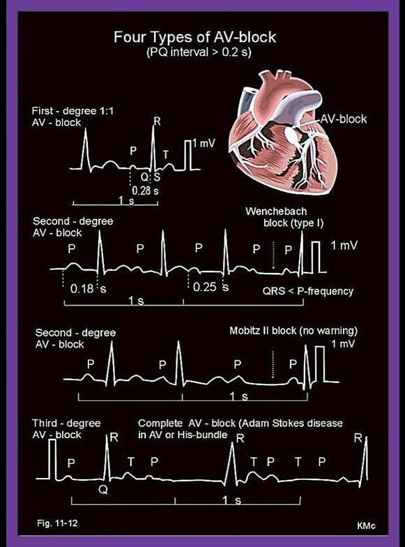
There are three basic types of AV nodal block:

  • First-degree AV block.
  • Second-degree AV block. Type I second-degree AV block (Mobitz I), also known as Wenckebach block. Type 2 second-degree AV block (Mobitz II) – due to a block in or below the bundle of His.
  • Third-degree AV block (complete heart block)

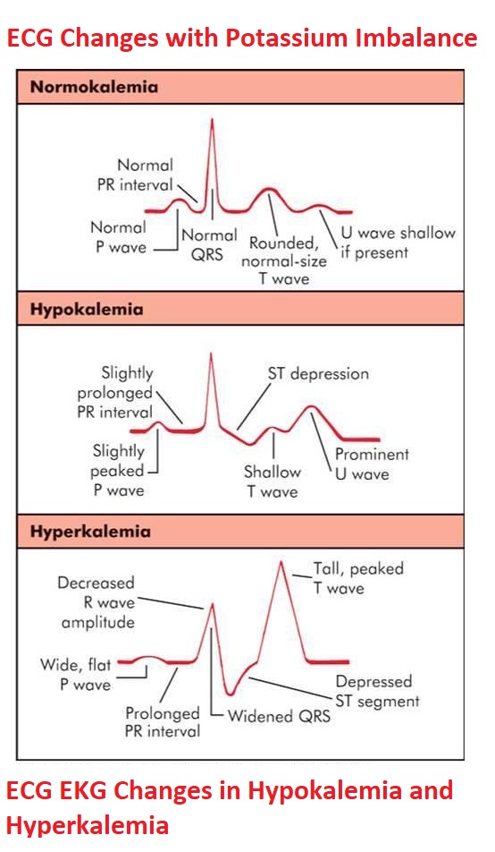
First-degree atrioventricular block (AV block), or PR prolongation, is a disease of the electrical conduction system of the heart in which the PR interval is lengthened beyond 0.20 seconds.

Type 2 Second-degree AV block, also known as “Mobitz II,” is almost always a disease of the distal conduction system (His-Purkinje System). Mobitz II heart block is characterized on a surface ECG by intermittently nonconducted P waves not preceded by PR prolongation and not followed by PR shortening.

Third-degree atrioventricular block (AV block), also known as complete heart block, is a medical condition in which the nerve impulse generated in the sinoatrial node (SA node) in the atrium of the heart does not propagate to the ventricle.

ECG Heart Block Rhythms - Four types of atrio-ventricular (AV)-block

Left Bundle Branch Block (LBBB) ECG Criteria

Left Bundle Branch Block (LBBB) ECG Criteria

The ECG criteria for a left bundle branch block include:

  • QRS duration greater than 120 milliseconds
  • Absence of Q wave in leads I, V5 and V6
  • Monomorphic R wave in I, V5 and V6
  • ST and T wave displacement opposite to the major deflection of the QRS complex

Left Bundle Branch Block (LBBB) ECG Criteria

A simple way to diagnose a left bundle branch in an ECG with a widened QRS complex (> 120 ms) would be to look at lead V1. If the QRS complex is widened and downwardly deflected in lead V1, a left bundle branch block is present. If the QRS complex is widened and upwardly deflected in lead V1, a right bundle branch block is present. The image below shows the typical findings of a left bundle branch block in the precordial ECG leads.