Right Bundle Branch Block (RBBB) Ecg Criteria

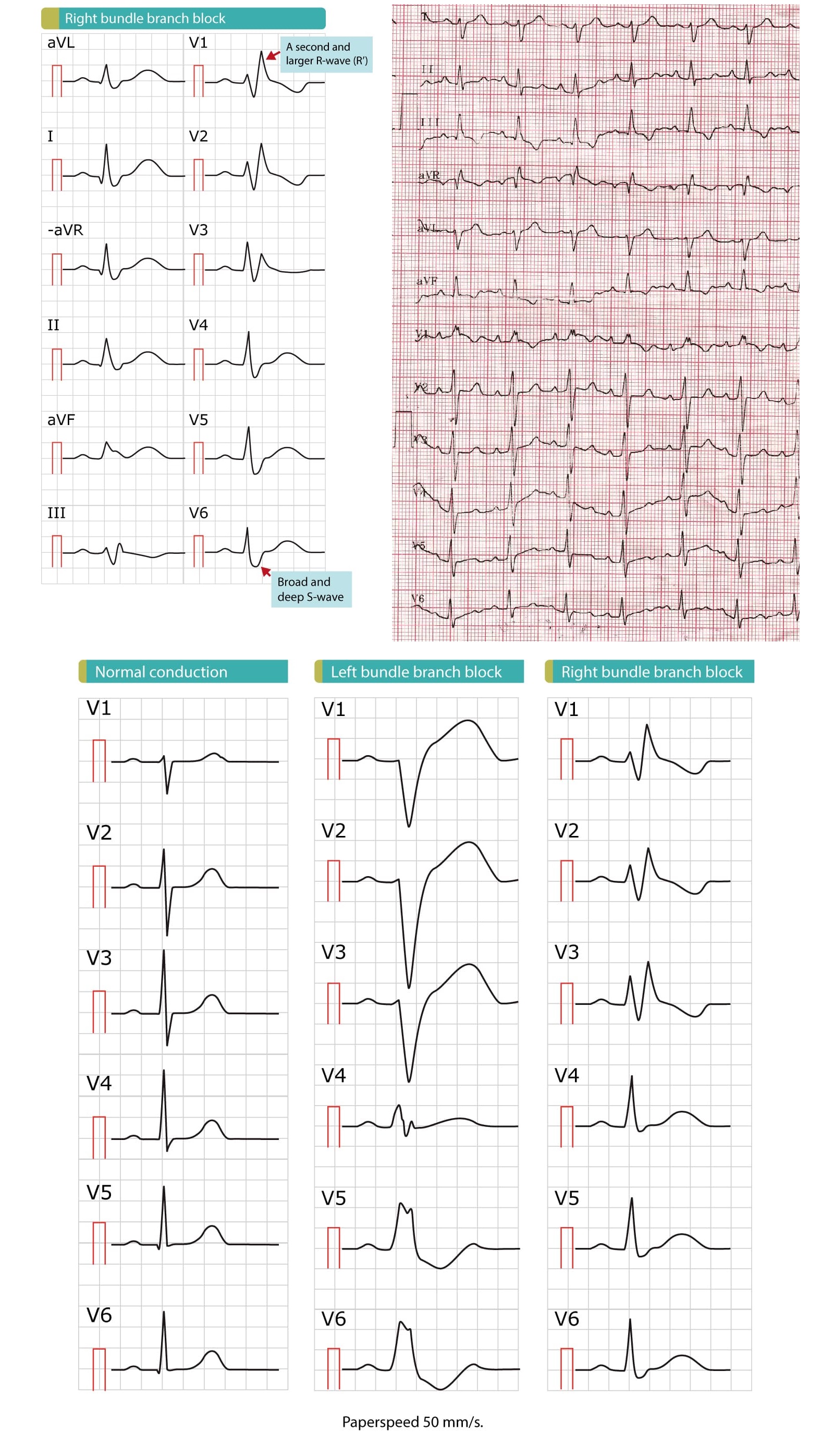
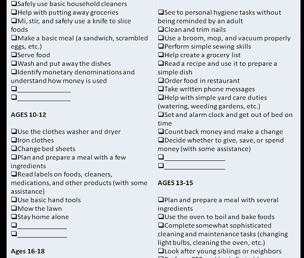
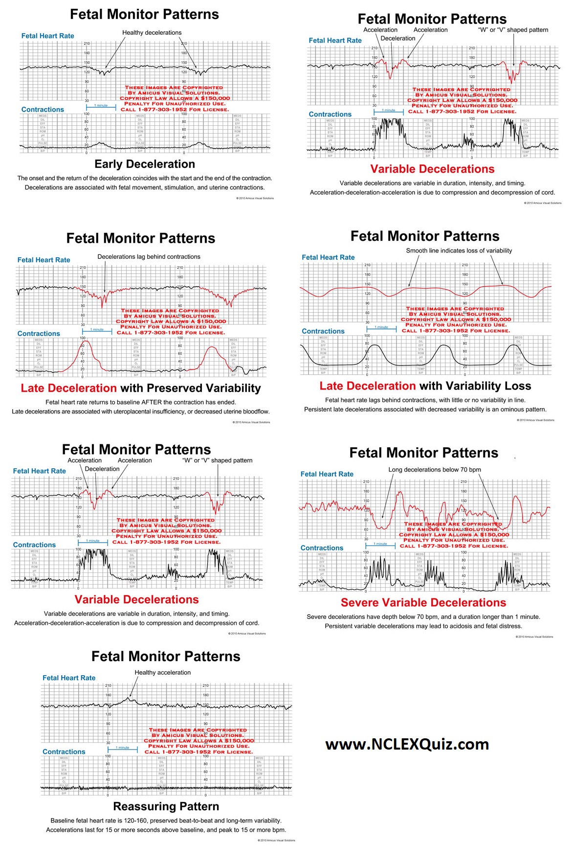
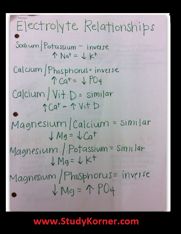
The ECG criteria for a right bundle branch block include the following: QRS duration greater than 120 milliseconds. rsR’ “bunny ear” pattern in the anterior precordial leads (leads V1-V3) Slurred S waves in leads I, aVL and frequently V5 and V6.

Right Bundle Branch Block (RBBB) Ecg Criteria

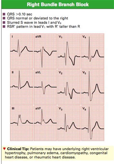
The ECG criteria for diagnosing right bundle branch block (RBBB) typically include the following:

QRS duration greater than 120 milliseconds: The QRS complex, which represents ventricular depolarization, is wider than normal due to delayed activation of the right ventricle.

rsR’ “bunny ear” pattern in the anterior precordial leads (leads V1-V3): The QRS complex shows an initial small r wave, followed by a larger R wave, and a second smaller r’ wave that gives the appearance of bunny ears.

Slurred S waves in leads I, aVL and frequently V5 and V6: The S wave, which represents initial negative deflection in the QRS complex, is prolonged and has a slurred appearance in the lateral leads I, aVL, and V5-V6.

Right Bundle Branch Block RBBB Ecg Criteria Definition

Comprehensive Checklist of Life Skills for Ages 2-18

Comprehensive Checklist of Life Skills for Ages 2-18

As you’re looking through the list, please remember that this is a guide. There’s no need to panic if you’re child isn’t straightening her bookshelves by seven. But hopefully it will give you a good idea of the age appropriate skills she’s ready to tackle along the way.

(Click on the image to download a larger, printable copy of the list.)

Comprehensive Checklist of Life Skills for Ages 2-18

Comprehensive Checklist of Life Skills for Ages 2-18

How to Memorize the H’s and T’s of ACLS – Certification Training for Nurses

SUDDEN CARDIAC ARREST, or SCA, is defined as “a sudden and unexpected pulseless condition attributed to cessation of cardiac mechanical activity”, according to the Sudden Cardiac Arrest Foundation. SCA is the leading cause of death in adults over the age of 40 in the United States, but fortunately is often reversible and can be treated with early intervention.

Understanding what protocols and actions to take when a patient is experiencing cardiac arrest is a crucial part of Advanced Cardiac Life Support (ACLS). They are complex, so practitioners and teachers have developed a mnemonic device, known as the “H’s and T’s of ACLS”, which explain the conditions associated with pre-arrest and cardiac arrest algorithms.

Because many cardiac arrest conditions are reversible, determining and treating the cause is a must for optimal patient care and outcome. Here are the H’s and T’s of ACLS from the latest American Heart Association standards and guidelines and AMC.

"T's"of ACLS Chart - ACLS Medical Training

"H's"of ACLS Chart - ACLS Medical Training

“T’s”of ACLS Chart – ACLS Certification Training for Nurses

What are the “T’s”?

"T's"of ACLS Chart - ACLS Medical Training

Tamponade (Cardiac)
Cardiac tamponade is a buildup of blood or fluid in the pericardial space, causing pressure on the heart, preventing ventricles from filling properly. Causes of tamponade include chest trauma, pericarditis and myocardial rupture.

Toxins
The ingestion of toxins is one of the most common causes of cardiac arrest, and occurs when an individual either intentionally or unintentionally overdoses on some type of medication, street drug or chemical exposure. One commonly seen sign of cardiac arrest due to toxins is a prolonged QT interval.

Tension Pneumothorax
Tension pneumothorax develops when there is a buildup of air in the pleural cavity, but there is no place for air to exit. The buildup of air causes a shift in mediastinum and venous return to the heart is blocked and can result in cardiac arrest. Some common causes of tension pneumothorax are chest trauma, iatrogenic and mechanical ventilation. Signs include unequal breathing sounds, JVD, ventilation issues and tracheal deviation.

Thrombosis (Pulmonary)
Thrombosis, or a pulmonary embolism, typically develops after a blood clot in another area of the body, and can lead to cardiac arrest in some instances. A pulmonary embolism is a blockage of the lung’s main artery, and is caused by blood clots.

Thrombosis (Coronary)
Coronary thrombosis is a blockage within the coronary artery or arteries due to clotted blood in the vessel. This prevents blood from properly flowing to the heart, and can cause cardiac arrest depending on the intensity and location of the blockage. Coronary thrombosis is caused by blood clots and myocardial infarction.

“H’s”of ACLS Chart – ACLS Certification Training for Nurses

What are the “H’s”?

"H's"of ACLS Chart - ACLS Medical Training

Hypovolemia
Hypovolemia is a common cause of cardiac arrest and develops from severe fluid or blood loss. This is typically a result of excessive sweating, vomiting, traumatic blood loss, severe burns or diarrhea.

Hypoxia or Hypoxemia
Hypoxia is an insufficiency in the amount of oxygen that reaches vital organs and tissues. It can occur from several reasons, including disorders of the lungs, drowning, fires, high altitudes, chemical or gas poisonings, etc.

Hydrogen Ion Excess (or Acidosis)
Hydrogen Ion Excess, or Acidosis, is either a respiratory or metabolic emergency that can lead to cardiac arrest that occurs when there are inadequate pH levels caused by too much acid in the body. Some causes of acidosis include lung disease, C02 buildup, DKA/AKA, liver failure, cancer, etc.

Hypokalemia and Hyperkalemia
Both hypokalemia and hyperkalemia are conditions in which potassium levels are too high or low to maintain normal contraction of the myocardium. When these levels of K+ are off, the individual is at high risk for cardiac arrest. A few causes of hypokalemia and hyperkalemia include use of diuretics, excessive vomiting, DKA, and kidney disease or failure. Hyperkalemia may be caused by diabetes, kidney disease or as a side effect of certain drugs.

Hypothermia
Hypothermia occurs when the body cannot keep itself warm and the core temperature drops below 30 degrees Celsius. Hypothermia can lead to cardiac arrest because cardiac output is decreased. Hypothermia ensues when an individual is exposed to extreme and/or extended exposure to cold weather or water temperatures.

Labeled Radiographic Anatomy & Differences Between Male & Female Pelvis

Labeled Radiographic Anatomy & Differences Between Male & Female Pelvis

Labeled Radiographic Anatomy & Differences Between Male & Female Pelvis
Labeled radiographic anatomy of the male (bottom image) and female (top image) pelvis.

The male pelvic inlet is narrower than the female pelvic inlet and is more heart shaped, whereas the female pelvic inlet is more circular and wider. In general, the male pelvis is more tall and narrow while the female pelvis is more broad and flattened. Additionally, the angle beneath the ischia bones (the pubic arch) forms a much more acute angle in the male (<90 degrees) while in the female the pubic arch forms an obtuse angle (>90 degrees), and the pubic arch is higher in the male. Additionally, the obturator foramen in the male are round, whereas in the female they are oval.

Male versus Female Pelvis – Labeled Radiographic Anatomy
Labeled female and male pelvis demonstrating the differences between the male and female pelvis.

Interpretation of the Electronic Fetal Heart Rate During Labor

Intrapartum Fetal Heart Rate Monitoring
The fetal heart rate tracing shows ALL of the following: Baseline FHR 110-160 BPM, moderate FHR variability, accelerations may be present or absent, no late or variable decelerations, may have early decelerations.

MATERNAL/CHILD HEALTH (OB): FETAL HEART TONES ACRONYM

VEAL CHOP
Variable decelerations…..Cord compression
Early decelerations………Head compression
Accelerations…………….OK, may need Oxygen
Late decelerations……….Placental Insufficiency

Interpretation of the Electronic Fetal Heart Rate During Labor

INTERVENTIONS
Variable decels → reposition mother to knee-chest position to get baby’s head off the cord OR use two fingers to lift the baby’s head off the cord until further interventions required
Early decels → sign that baby is descending into the pelvis, monitor as needed
Accelerations → reassuring (normal) sign; last for 15+ seconds and peaks 15+ beats/min
Late decels → worrisome sign; reposition mother, administer IV fluids and anticipate discontinuing/decreasing Oxytocin or administering a tocolytic to decrease contractions

Cardiac Enzymes A.K.A. ACP (Acute Cardiac Profile)

Cardiac Enzymes A.K.A. ACP (Acute Cardiac Profile)

Creatine Phosphokinase (CPK) Test

In a healthy adult, the serum CPK level varies with a number of factors (gender, race and activity), but normal range is 26 to 192 U/L (units per liter). Higher amounts of serum CPK can indicate muscle damage due to chronic disease or acute muscle injury.

Cardiac Enzymes A.K.A. ACP (Acute Cardiac Profile)

This test may be used to:

Diagnose heart attack
Evaluate cause of chest pain
Determine if or how badly a muscle is damaged
Detect dermatomyositis, polymyositis, and other muscle diseases
Tell the difference between malignant hyperthermia and postoperative infection