Disease Specific Isolation Recommendation Cheatsheet
- Standard Precautions
- Contact Precautions
- Droplet Precautions
- Airborne Precautions

Disease Specific Isolation Recommendation Cheatsheet
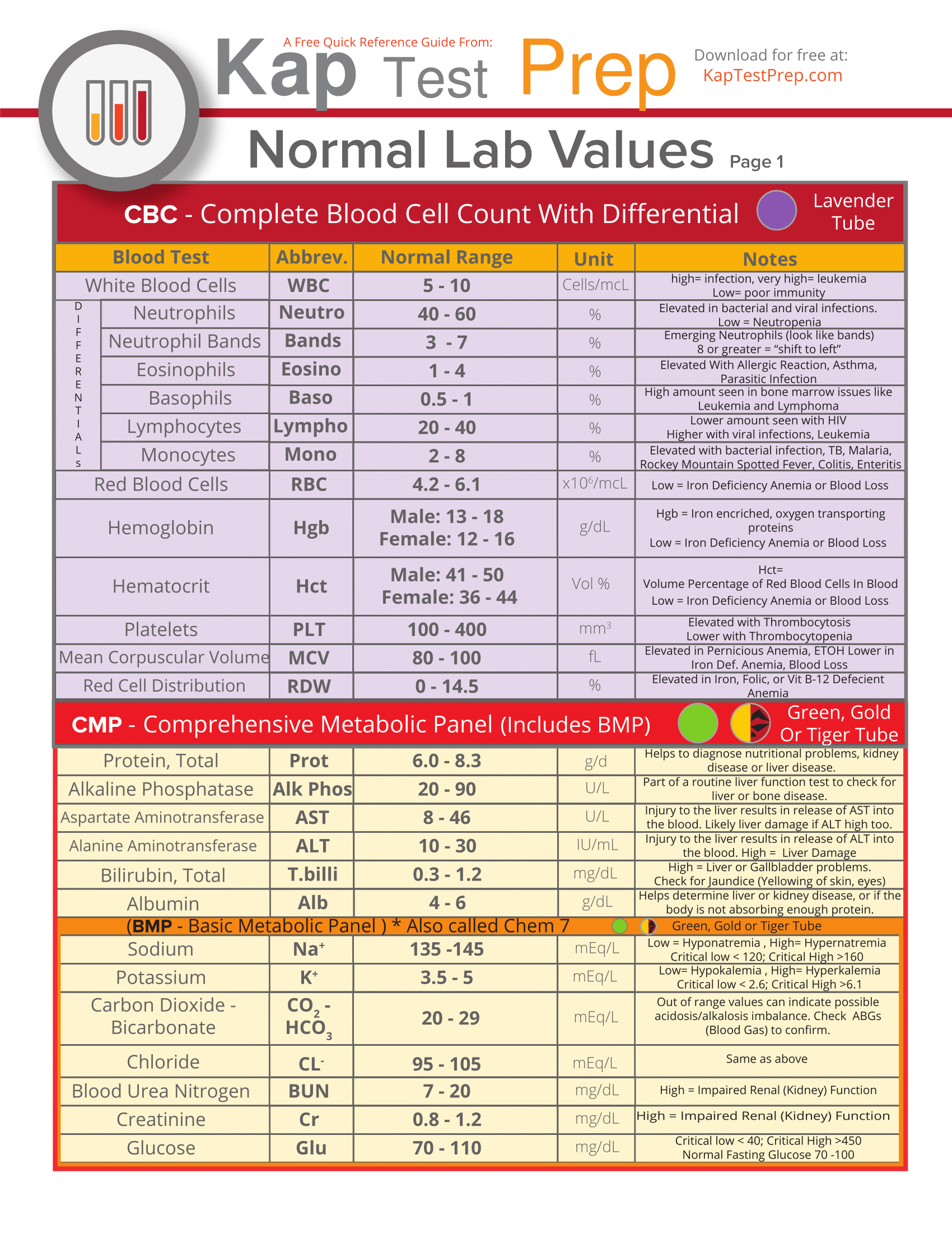
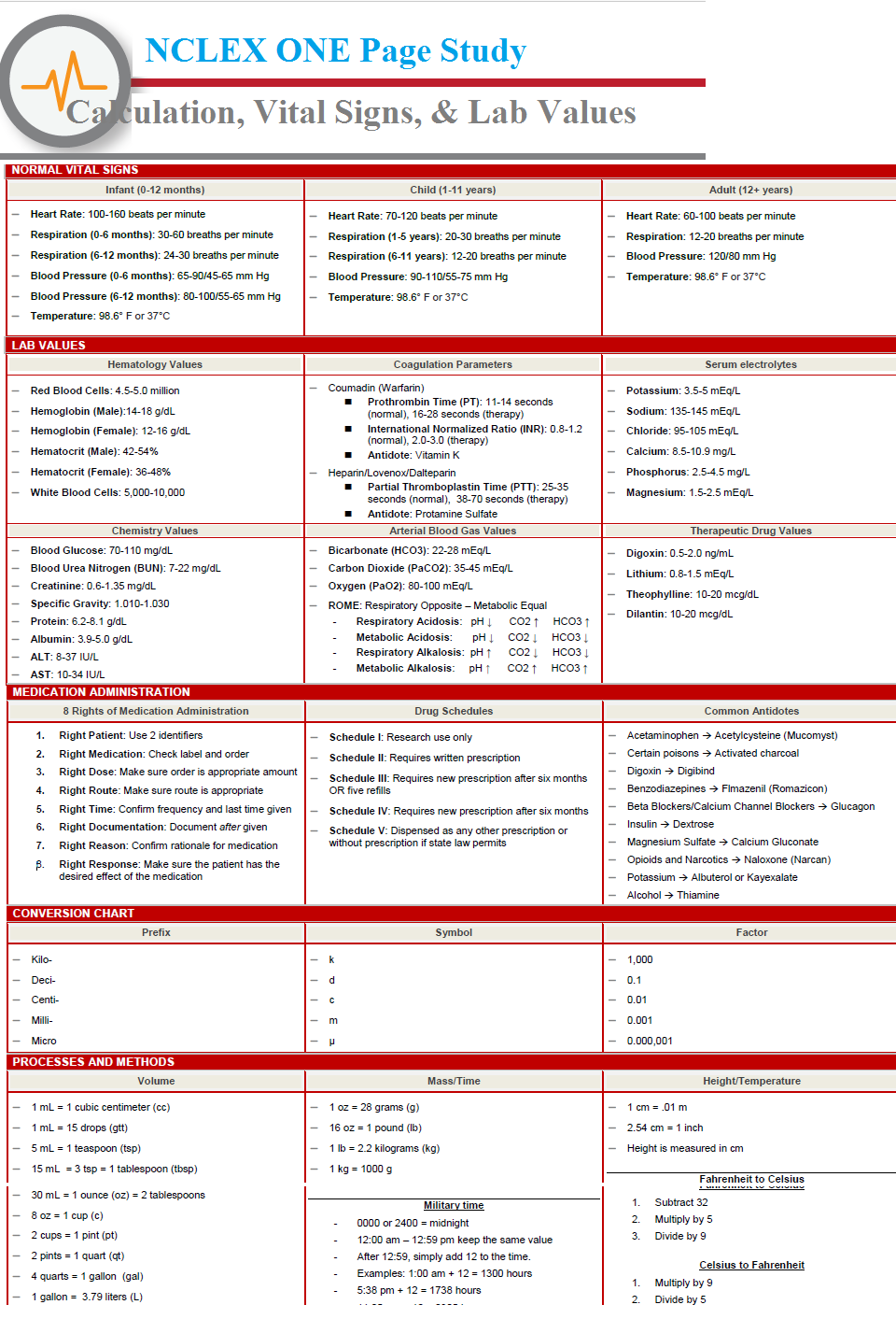
Laboratory values provide healthcare professionals with essential clues about the patient’s condition and the medical interventions needed for his full recovery. As nurses are the first-line responders to the healthcare needs of patients, we should always be familiar with the common laboratory values and how to interpret them.
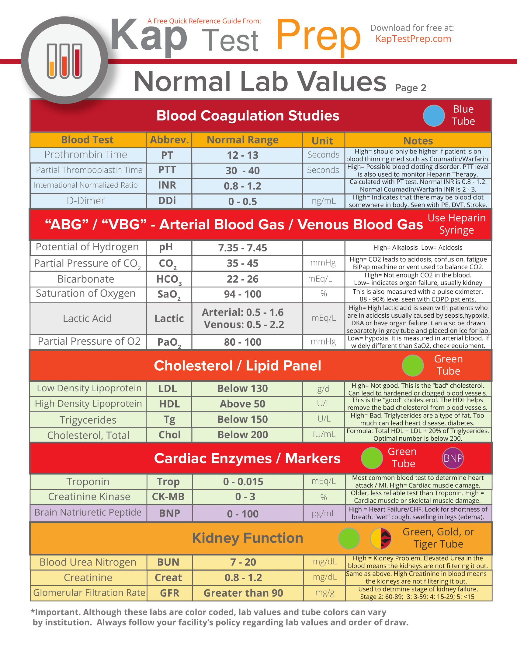
Laboratory values provide healthcare professionals with essential clues about the patient’s condition and the medical interventions needed for his full recovery. As nurses are the first-line responders to the healthcare needs of patients, we should always be familiar with the common laboratory values and how to interpret them.
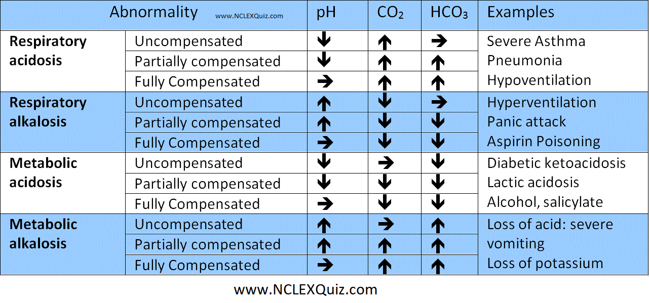
The body normally controls the pH of blood within a tight range. One must always remember that pH is a logarithmic scale and so a change from 8 to 7 is a ten-fold increase in H+ concentration. A normal intracellular pH is required for the functioning of many enzyme systems. When blood becomes profoundly acidotic (pH<7) then cellular function becomes impossible and death ensues. There are a lot of texts available describing the causes of the respiratory and metabolic acidosis and alkalosis. However the best way to learn how to interpret blood gases is to practice.
The Advanced Paediatric Life Support Course describes a simple three step system to interpreting paediatric blood gases (do not feel you have to use this method if you already have a good system). You should always keep the Henderson-Hasselbach equation in the back of your mind whilst doing this.
Henderson-Hasselbach equation
CO2 + H2O = H2CO3 = H+ + HCO3-
Step 1 – What is the pH? Is there an acidosis or alkalosis?
Step 2 – What is the CO2?
If the CO2 provides a cause of the abnormal pH i.e low pH with a high CO2 (acidosis) or high pH with low CO22 (alkalosis), then the overall picture is RESPIRATORY.
If the CO2 does not provide a cause of the pH, then it is metabolic (possibly compensating).
Step 3 – Confirm your findings by looking at the base excess or bicarbonate.
If the base excess provides a cause for the abnormal pH, i.e. low pH with a negative base excess (acidosis) OR high pH with positive base excess (alkalosis), the overall picture is METABOLIC.
If the base excess does not provide a cause for the pH, it’s compensating for a respiratory abnormality..
Keep this travel nurse packing list on hand when preparing for your assignments, and you’ll greatly enhance the chances of making your transition a smooth and relatively low-stress one.

NCLEX One Page Study is a Short time Learning. following are the Main Points of NCLEX One Page Study.
Isolation Precaution Cheat Sheet. This will help nursing students.

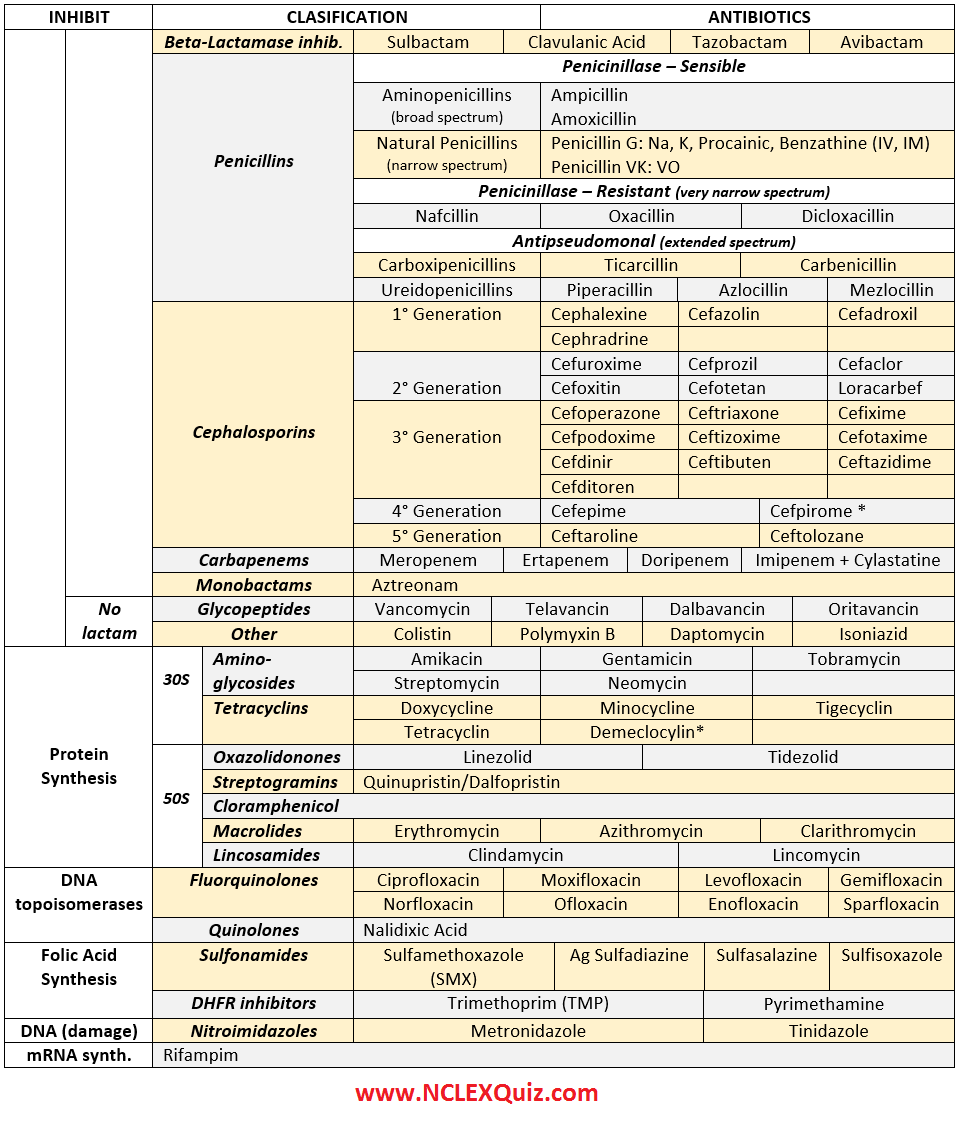
ANTIBIOTICS CHEAT SHEET 🙂
Also, REMEMBER!!!!
* Sulfonamides compete for albumin with:
Bilirrubin: given in 2°,3°T, high risk or indirect hyperBb and kernicterus in premies
Warfarin: increases toxicity: bleeding
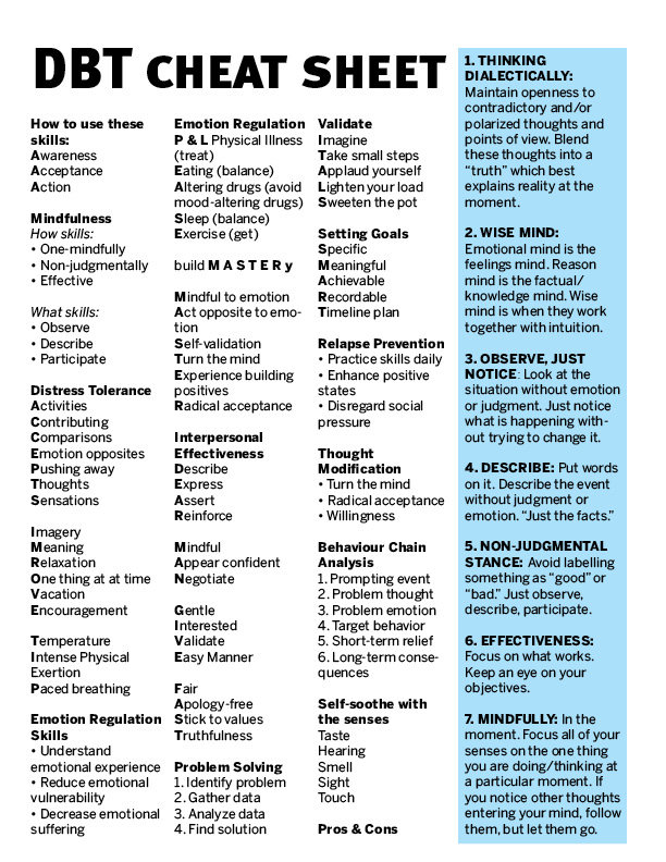
Dialectical Behavior Therapy (DBT) is a cognitive behavioral treatment that was originally developed to treat chronically suicidal individuals diagnosed with borderline personality disorder (BPD) and it is now recognized as the gold standard psychological treatment for this population. In addition, research has shown that it is effective in treating a wide range of other disorders such as substance dependence, depression, post-traumatic stress disorder (PTSD), and eating disorders.
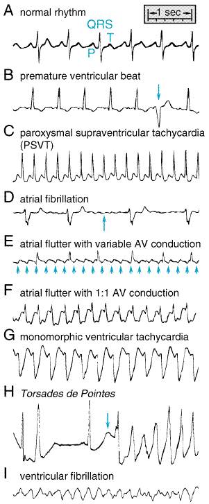
ECGs Showing Normal and Abnormal Cardiac Rhythms. The P, QRS, and T waves in normal sinus rhythm are shown in panel A. Panel B shows a premature beat arising in the ventricle (arrow). Paroxysmal supraventricular tachycardia (PSVT) is shown in panel C; this is most likely reentry utilizing an accessory pathway or reentry within or near the AV node. In atrial fibrillation (panel D), there are no P waves and the QRS complexes occur irregularly (and at a slow rate in this example); electrical activity between QRS complexes shows small undulations (arrow), corresponding to fibrillatory activity in the atria. In atrial flutter (panel E), the atria beat rapidly, approximately 250 beats per minute (arrows) in this example, and the ventricular rate is irregular. If a drug that slows the rate of atrial flutter is administered, 1:1 atrioventricular conduction (panel F) can occur. In monomorphic ventricular tachycardia (VT, panel G), identical, wide QRS complexes occur at a regular rate, 180 per minute. The electrocardiographic features of the torsades de pointes syndrome (panel H) include a very long QT interval (>600 ms in this example, arrow) and ventricular tachycardia in which each successive beat has a different morphology (polymorphic VT). Panel I shows the disorganized electrical activity characteristic of ventricular fibrillation.