Chest X-Ray Lung disease Four-Pattern Approach
On a chest x-ray lung abnormalities will either present as areas of increased density or as areas of decreased density.
Lung abnormalities with an increased density – also called opacities – are the most common.
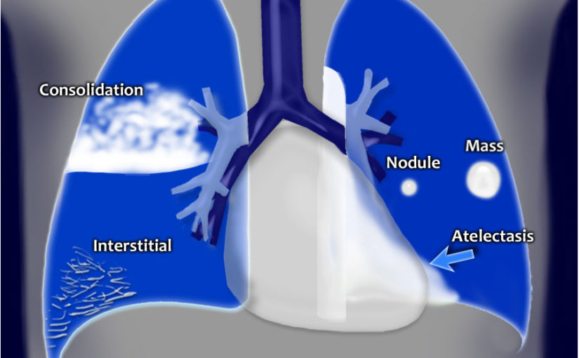
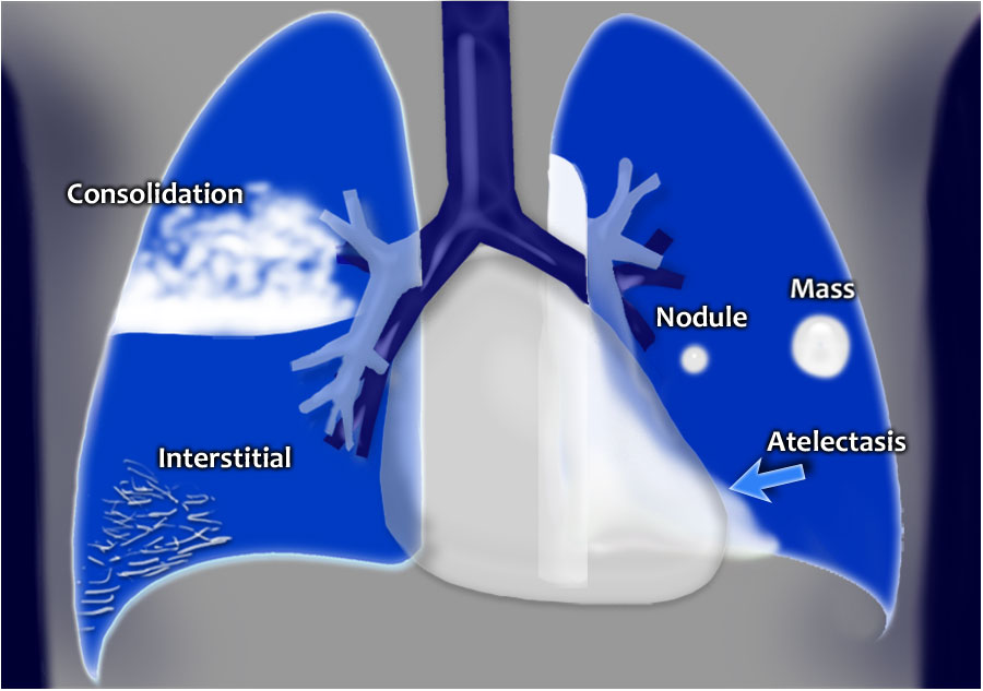
A practical approach is to divide these into four patterns:
- Consolidation
- Interstitial
- Nodules or masses
- Atelectasis
4-Pattern approach
Whenever you see an area of increased density within the lung, it must be the result of one of these four patterns.
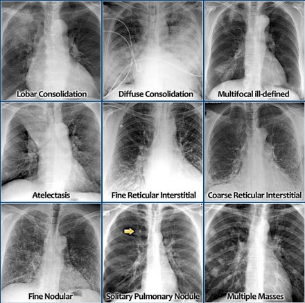
Consolidation – any pathologic process that fills the alveoli with fluid, pus, blood, cells (including tumor cells) or other substances resulting in lobar, diffuse or multifocal ill-defined opacities.
Interstitial – involvement of the supporting tissue of the lung parenchyma resulting in fine or coarse reticular opacities or small nodules.
Nodule or mass – any space occupying lesion either solitary or multiple.
Atelectasis – collapse of a part of the lung due to a decrease in the amount of air in the alveoli resulting in volume loss and increased density.
Here are the most common examples of these four patterns on a chest x-ray
Consolidation
Lobar consolidation
Diffuse consolidation
Multifocal ill-defined consolidations
Interstitial
Reticular interstitial opacities
Fine Nodular interstitial opacities
Nodule or mass
Solitary Pulmonary Nodule
Multiple Masses
Atelectasis