NCLEX Cheat Sheets: Phases of the cardiac cycle PQRST Wave for Nursing Students

NCLEX Cheat Sheets: Phases of the cardiac cycle for Nursing Students

cardiac cycle pqrst wave
cardiac cycle in physiology
Cardiac cycle of heart
cardiac cycle time

5 Steps of Cardiac Cycle
The cardiac cycle is essentially split into two phases, systole (the contraction phase) and diastole (the relaxation phase). Each of these is then further divided into an atrial and ventricular component.

Passive Ventricular Filling.
Passive Ventricular Filling. AV valve is open, the SL valve is closed.
Atrial contraction and emptying. AV valve will close, SL valve stays closed.
Isovolumic contraction.
Ventricular contraction and ejection.
Isovolumic relaxation.

The cardiac cycle comprises a complete relaxation and contraction of both the atria and ventricles, and lasts approximately 0.8 seconds.

NCLEX Cheat Sheets: Phases of the cardiac cycle PQRST Wave for Nursing Students

Tetralogy of Fallot Rapid Review For Nursing Students

Tetralogy of Fallot Rapid Review For Nursing Students

Tetralogy of Fallot

  • Patient with a history of episodes of cyanosis (tet spells) and squatting for relief
  • PE will show pulmonic stenosis, right ventricular hypertrophy, overriding aorta, VSD
  • CXR will show “boot-shaped” heart
  • Comments: Most common cyanotic congenital heart disease
  • Mnemonic: PROVePulmonic stenosis, Right ventricular hypertrophy, Overriding aorta, VSD

Tetralogy of Fallot Rapid Review For Nursing Students

Multiple Sclerosis Nursing: Signs and Symptoms

Multiple Sclerosis Nursing: Signs and Symptoms

Multiple sclerosis – causes, symptoms, diagnosis, treatment, pathology

Multiple sclerosis, or MS, is a long-lasting disease that can affect your brain, spinal cord, and the optic nerves in your eyes.

common symptoms of MS are:

Clumsiness or weakness
Difficulty walking or maintaining balance
Dizziness or vertigo
Eye problems such as double vision or uncontrolled eye movements
Fatigue
Muscle spasms
Problems with bladder or bowel control
Stiffness
Tremors
Tingling or numbness
Depression or emotional changes
Mild intellectual change such as memory problems
Sexual problems such as difficulty reaching orgasm, lack of vaginal sensation or erectile dysfunction
Symptoms usually appear in people between 20 and 40 years of age.

Multiple Sclerosis Nursing: Signs and Symptoms

Nursing Notes: Difference between Atelectasis and Pneumothorax

Nursing Study Tips: Difference between Atelectasis and Pneumothorax
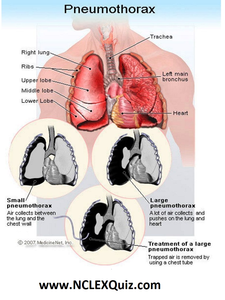
A Pneumothorax takes place when there is a tear in the lung and air builds up between the outside of your lungs and the inside of your chest.

Atelectasis is when one or more lobes (sections) of the lung collapse because of a blockage or pressure inside or outside the bronchial tubes in the lungs. The blockage causes air to become trapped, creating a sensation of shortness of breath. Blockages can be caused by:

1. Mucus plug
2. Tumor
3. Inhaled foreign object

A pneumothorax occurs when there is air or gas in the lung pleural cavity compressing some of the lung tissue. There are varying degrees of a pneumothorax (PTX) as some are mild and require only monitoring and others can be life threatening.

If it is mild, he may be monitored by his primary care physician or a pulmonologist (lung specialist), but if it is more severe, he may be admitted to the hospital. There are various causes for a pneumothorax, including trauma and spontaneous development.

A life threatening condition can occur when a tension pneumothorax develops and displaces a patient’s mediastinal structures (including heart and trachea). This can cause significant respiratory and cardiovascular compromise and requires emergent evaluation and treatment.

Nursing Notes: Difference between Atelectasis and Pneumothorax

Pulmonary Embolism Pathophysiology Nursing

Pulmonary Embolism Pathophysiology Nursing

Pulmonary embolism (PE) occurs when a pulmonary artery becomes blocked—usually by a blood clot that has broken free from its site of origin and embolized or migrated to the lungs. If misdiagnosed, unrecognized, or untreated, PE can cause death quickly—within just an hour. It’s fatal in up to 26% of cases.

Massive PE, defined as causing 50% or more occlusion of the pulmonary capillary bed, can result in obstructive shock with systemic hypoperfusion (low cardiac output and acute pulmonary hypertension with right ventricular failure). It must be remedied immediately to save the patient’s life.

Pathophysiology
The usual underlying cause of PE is deep vein thrombosis (DVT) in a lower extremity, the pelvis, or even an upper extremity. DVT, in turn, typically results from one or more of these conditions: venous stasis, blood hypercoagulability (increased clotting), or endovascular damage. Known collectively as Virchow’s triad, these conditions can stem from a range of situations.

Pulmonary Embolism Pathophysiology Nursing

Nursing Diagnosis for Myocarditis

Myocarditis Disease with Causes, Symptom and Nursing Intervention

Myocarditis disease or Inflammatory cardiomyopathy is an inflammation of the myocardium, the middle layer of the heart wall.

The following diagnostic tests can help you determine if a patient has myocarditis or another condition. A chest X-ray may show an enlarged heart with evidence of heart failure, such as prominent blood vessels or fluid within the lungs. An ECG may show arrhythmias, as well as ST-segment and T-wave abnormalities.

Myocarditis Disease with Causes, Symptom and Nursing Intervention